Learn why calcific tendonitis is painful and explore treatment options with expert insights from Mr. Toby Baring.


Calcific tendonitis is a distressing condition that causes significant, often debilitating pain through the accumulation of calcium deposits within the tendons of the shoulder. To understand why calcific tendonitis is so painful, it helps to examine the role of calcium deposits, the resulting inflammation, and their combined impact on tendon function.
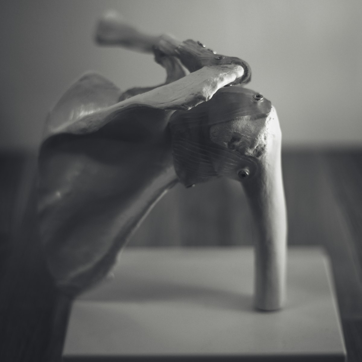
Calcific tendonitis is characterised by the deposition of calcium salts in tendons, with a particular predilection for the rotator cuff in the shoulder. These calcium deposits can lead to intense inflammation and discomfort, severely impairing the mobility and functionality of the affected tendon.
Patients with calcific tendonitis typically experience severe pain, which may start abruptly and be severely debilitating. The pain often intensifies with arm movement or during the night, significantly affecting sleep quality. Other symptoms include a noticeable reduction in range of motion, tenderness upon touch, and visible swelling around the affected area. A person with severe calcific tendonitis in the shoulder may find it difficult to lift objects, perform overhead tasks, or carry out simple daily activities like dressing.
The formation of calcium deposits is the principal source of pain in calcific tendonitis. These deposits are rigid and irritate the surrounding soft tissues, especially the bursa, leading to significant discomfort. The pain often intensifies as the body attempts to break down and absorb these calcium deposits, a process known as resorption. This resorption phase can be prolonged and notably painful, as it triggers an inflammatory response that further compounds the discomfort.
Inflammation plays a central role in the painful nature of calcific tendonitis. When the immune system targets the calcium deposits, it causes the surrounding tissues to become inflamed. This inflammation not only precipitates pain but also leads to stiffness and swelling, which can severely restrict movement. During a flare-up, a person might experience real difficulty reaching overhead or behind their back — movements that are fundamental to many everyday tasks.
A thorough clinical examination is vital for diagnosing calcific tendonitis. Orthopaedic specialists conduct a detailed assessment of the patient's symptoms and medical history, combined with a physical examination to evaluate range of motion and identify the precise source of pain.
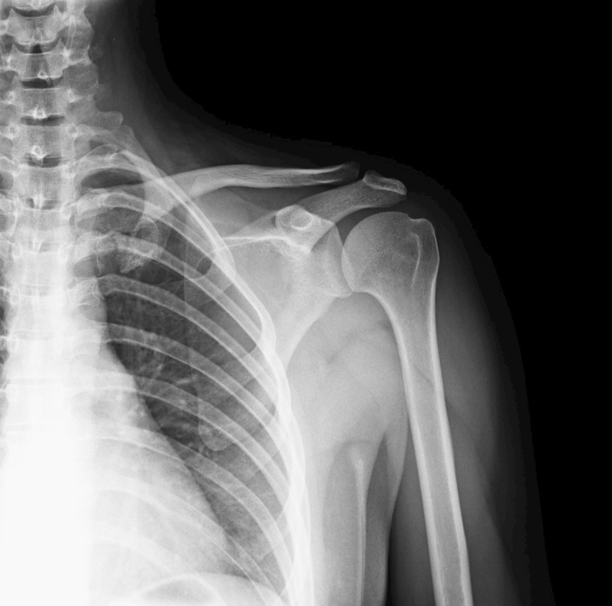
Advanced imaging techniques are indispensable for confirming a diagnosis of calcific tendonitis. X-rays are frequently employed to identify calcium deposits, offering a clear view of their size and location. Ultrasound and MRI scans provide further detail by illustrating the condition of the tendons and surrounding tissues, allowing clinicians to assess the extent and severity of the condition accurately. These imaging tools are particularly useful for planning treatment strategies, as they help differentiate calcific tendonitis from other similar conditions such as rotator cuff tears or subacromial bursitis.
A differential diagnosis is needed to distinguish calcific tendonitis from conditions with overlapping symptoms, such as rotator cuff tears, bursitis, or frozen shoulder. Accurate identification is fundamental to implementing the most effective treatment plan. Understanding the distinct characteristics of calcific tendonitis, particularly the presence of calcium deposits visible on imaging, ensures that management strategies are tailored to address the specific challenges of the condition.
Non-surgical treatments form the cornerstone of initial management for calcific tendonitis. These treatments aim to alleviate pain, reduce inflammation, and restore functionality. Key non-surgical options include:
Surgical intervention may be considered when non-surgical treatments fail to provide sufficient relief. Surgical options are targeted at removing the calcium deposits and restoring tendon function. These include:

The recovery process from calcific tendonitis varies depending on the chosen treatment approach. Post-treatment rehabilitation programmes are integral to a successful outcome, focusing on progressive strengthening and stretching exercises to restore full function and reduce the risk of recurrence. A structured rehabilitation plan may involve gradually increasing the intensity and complexity of exercises, allowing the tendon to heal while rebuilding strength. Ongoing monitoring by a physical therapist ensures exercises are performed correctly and that recovery progresses at an appropriate pace.
Why is calcific tendonitis so painful?
Calcific tendonitis is painful due to the accumulation of calcium deposits in the tendons, leading to inflammation and pressure on surrounding tissues.
What is the pain scale for calcific tendonitis?
The pain scale for calcific tendonitis can vary, but it often ranges from moderate to severe, depending on the extent of calcium deposits and inflammation.
How long does it take for calcific tendonitis to reabsorb?
Calcific tendonitis can take several weeks to months to reabsorb naturally, but this varies based on individual cases and treatment approaches.
Can you make calcific tendonitis worse?
Yes, certain activities or lack of treatment can exacerbate calcific tendonitis, increasing pain and prolonging recovery.
If you're experiencing symptoms of calcific tendonitis, contact us today to schedule a consultation with Mr. Toby Baring.